
Laboratory. Source: Murat Kizaibek via Wikimedia Commons.
Highlights
- Immunoblotting detects proteins involved in infection and disease.
- It reveals how viruses interact with the immune system.
- These discoveries support diagnostics, therapeutics, and vaccine development.
When most people think about public health, they picture vaccines, hospitals, or disease outbreaks. What they do not picture are the laboratory techniques that help scientists understand the pathogens behind those headlines.
Throughout my PhD, I have used immunoblotting extensively to study how host proteins interact with viruses and regulate antiviral immune responses. Some of my favorite moments in the lab came from developing a blot and seeing a protein band appear exactly where we predicted it would.
To many people, a Western blot looks like a series of dark bands on a membrane. To scientists, those bands reveal whether immune pathways are activated, treatments are working, or proteins are helping or hindering infection.
Importantly, although immunoblotting is performed at the lab bench, the discoveries it enables can ultimately shape diagnostics, therapeutics, vaccine development, and public health.
So, what exactly is immunoblotting, and why does it matter?
What Is Immunoblotting?
Immunoblotting is a laboratory technique used to detect specific proteins within a biological sample.
Proteins are the workhorses of the cell. They help viruses replicate, allow immune cells to communicate, and control countless biological processes.
By measuring the presence or abundance of particular proteins, scientists can learn what is happening inside infected or diseased cells.
How Immunoblotting Works

Visual representation of an SDS-PAGE gel. Source: Marta Ferreira via Wikimedia Commons.
First, scientists break open cells or tissues to extract their proteins. The proteins are then mixed with SDS sample buffer and heated. SDS is an essential ingredient because it unfolds (denatures) proteins and gives them a uniform negative charge. This negative charge ensures they migrate according to size rather than their natural shape or charge.
The samples are loaded into a polyacrylamide gel for gel electrophoresis. When an electric current is applied, the negatively charged proteins move toward the positive electrode. Smaller proteins migrate through the gel faster than larger ones, separating the proteins by molecular weight.
Next, the proteins are transferred from the gel onto a nitrocellulose or PVDF membrane using an electric current. During this step, the gel and membrane are immersed in transfer buffer, which conducts electricity, maintains protein stability, and helps proteins efficiently bind to the membrane while preserving their separation pattern.
The membrane is then blocked to prevent nonspecific antibody binding before being incubated with a primary antibody that recognizes the target protein. A secondary antibody, linked to an enzyme or fluorescent tag, binds the primary antibody and generates a detectable signal.
When the membrane is imaged, the target protein appears as a distinct band. Its position indicates the protein’s approximate size, while its intensity reflects its relative abundance, allowing researchers to compare protein expression across different samples and experimental conditions.
Why Proteins Matter in Infectious Disease Research
Viruses may carry genetic information, but proteins are ultimately responsible for many of the biological effects we observe during infection.
When a virus enters a cell, both viral and host proteins change dramatically. Some proteins help the virus replicate. Others are activated as part of the body’s defense system.
Immunoblotting allows scientists to monitor these changes in real time.
For example, researchers can measure whether an antiviral protein increases after infection, determine whether an immune signaling pathway has been activated, or evaluate whether an experimental drug successfully blocks a viral protein.
These insights help scientists understand why infections occur, how diseases progress, and what interventions may be effective.
Immunoblotting and the Immune Response
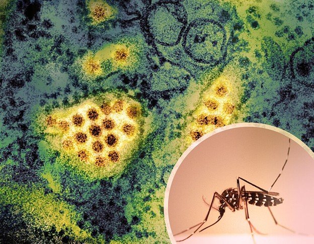
Aedes mosquito carrying dengue virus. Source: NIAID via Wikimedia Commons.
One of the most valuable applications of immunoblotting is studying the immune system.
When cells detect a virus, they activate signaling pathways that trigger the production of antiviral molecules. These pathways rely on proteins that switch on and off through a process known as phosphorylation.
Using immunoblotting, scientists can determine whether these proteins are activated during infection.
This information helps answer important questions:
- Is the immune system recognizing the virus?
- Is the virus blocking immune defenses?
- Are antiviral treatments restoring immune activity?
- Why do some infections cause severe disease while others remain mild?
By tracking these molecular events, researchers gain a deeper understanding of host-pathogen interactions.
How Immunoblotting Supports Public Health
Although immunoblotting is performed at the laboratory bench, its impact extends far beyond the research environment.
Many discoveries that shape public health policies begin with experiments designed to understand proteins and cellular pathways.
Immunoblotting has contributed to:
- Identifying proteins involved in viral replication
- Understanding mechanisms of antiviral immunity
- Validating potential drug targets
- Evaluating vaccine-induced immune responses
- Investigating emerging infectious diseases
The technique has been used extensively in research on viruses such as HIV, influenza, SARS-CoV-2, dengue virus, and measles virus.
By helping scientists understand how pathogens interact with human cells, immunoblotting provides the foundational knowledge needed to develop new diagnostics, therapeutics, and prevention strategies.
Strengths and Limitations
Like any scientific tool, immunoblotting has strengths and limitations.
One major advantage is specificity. Researchers can detect a single protein among thousands present in a sample. The technique is also relatively affordable and widely accessible, making it a staple of research laboratories worldwide.
However, immunoblotting is not perfect. Results depend heavily on antibody quality, proper controls, and careful experimental design. It also measures proteins in bulk populations of cells, meaning it cannot reveal what is happening within individual cells.
For this reason, scientists often combine immunoblotting with other techniques such as microscopy, flow cytometry, PCR, and next-generation sequencing.
Takeaway

Patient attended at the San Juan de Dios Hospital in Guatemala. Source: World Bank Photo via Flickr.
Behind every public health breakthrough lies a collection of laboratory tools that help scientists understand disease at the molecular level.
Immunoblotting may not generate headlines, but it remains one of the most powerful methods for studying how infections alter the proteins that govern cellular function. From uncovering antiviral defenses to identifying new therapeutic targets, this technique continues to shape our understanding of infectious diseases and improve public health outcomes around the world.
Subscribe to Pathogenos
Curious about the science behind infectious disease research?
The Inside the Lab series explores the techniques, technologies, and discoveries that drive modern public health. Subscribe to Pathogenos for evidence-based insights on pathogens, outbreaks, vaccines, and the laboratory tools used to understand them.


.jpg)